Bassett Collection of Stereoscopic Images of Human Anatomy
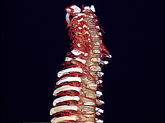
Arteries of vertebral column of one
Year-old infant - Arteries of cervicothoracic part of vertebral column, right anterolateral view
Image #211-1
KEYWORDS: Bones joints cartilage, Cervical region, Thoracic region, Vasculature, Vertebral column.
Creative Commons
Stanford holds the copyright to the David L. Bassett anatomical images and has assigned Creative Commons license Attribution-Share Alike 4.0 International to all of the images.

For additional information regarding use and permissions, please contact Dr. Drew Bourn at dbourn@stanford.edu.
|

| ||||||||||||||||||||||||||||||||||

|
|