Bassett Collection of Stereoscopic Images of Human Anatomy
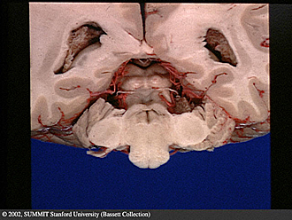
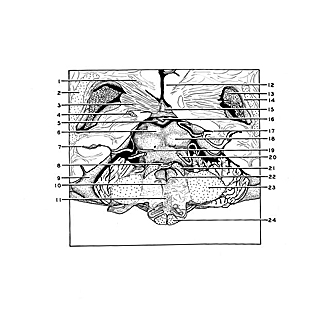
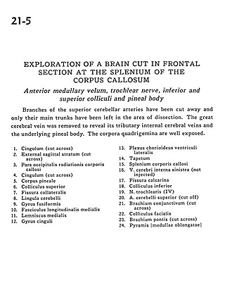
Exploration of a brain cut in frontal section at the splenium of the corpus callosum
Anterior medullary velum, trochlear nerve, inferior and superior colliculi and pineal body
Image #21-5
KEYWORDS: Brain, Diencephalon, Medulla, Telencephalon, Vasculature.
Creative Commons
Stanford holds the copyright to the David L. Bassett anatomical images and has assigned Creative Commons license Attribution-Share Alike 4.0 International to all of the images.

For additional information regarding use and permissions, please contact Dr. Drew Bourn at dbourn@stanford.edu.
|
| ||||||||||||||||||||||||||||||||||||||||||||||||||||
|
|