Bassett Collection of Stereoscopic Images of Human Anatomy
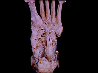
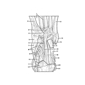
Joints of left ankle and foot
Ligaments of planar aspect of foot
Image #202-5
KEYWORDS: Ankle, Bones joints cartilage, Foot and toes, Muscles and tendons.
Creative Commons
Stanford holds the copyright to the David L. Bassett anatomical images and has assigned Creative Commons license Attribution-Share Alike 4.0 International to all of the images.

For additional information regarding use and permissions, please contact Dr. Drew Bourn at dbourn@stanford.edu.
|
| ||||||||||||||||||||||||||||||||||||||||||||||

|
|