Bassett Collection of Stereoscopic Images of Human Anatomy
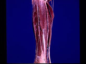
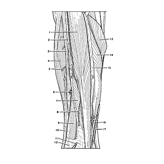
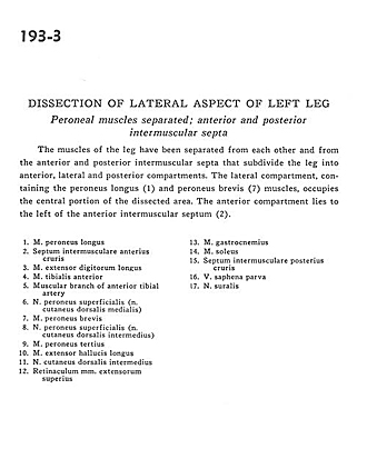
Dissection of lateral aspect of left leg
Peroneal muscles separated; anterior and posterior intermuscular septa
Image #193-3
KEYWORDS: Leg, Muscles and tendons.
Creative Commons
Stanford holds the copyright to the David L. Bassett anatomical images and has assigned Creative Commons license Attribution-Share Alike 4.0 International to all of the images.

For additional information regarding use and permissions, please contact Dr. Drew Bourn at dbourn@stanford.edu.
|
| ||||||||||||||||||||||||||||||||||||||
|
|