Bassett Collection of Stereoscopic Images of Human Anatomy
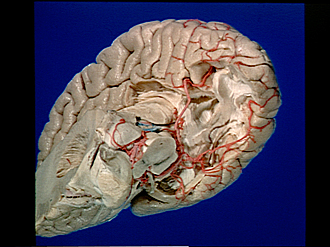
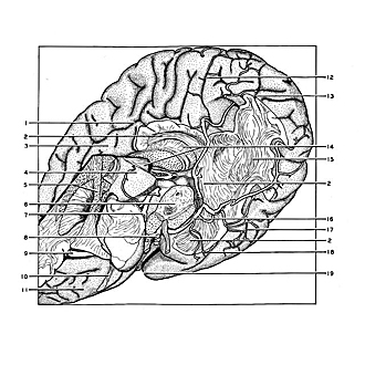
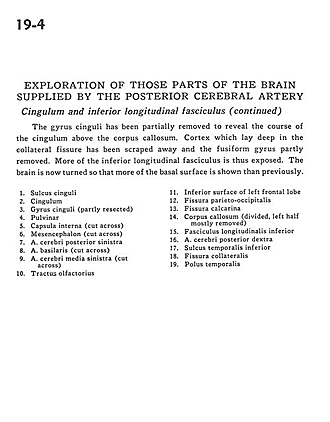
Exploration of those parts of the brain supplied by the posterior cerebral artery
Cingulum and inferior longitudinal fasciculus (continued)
Image #19-4
KEYWORDS: Brain, Telencephalon, Vasculature.
Creative Commons
Stanford holds the copyright to the David L. Bassett anatomical images and has assigned Creative Commons license Attribution-Share Alike 4.0 International to all of the images.

For additional information regarding use and permissions, please contact Dr. Drew Bourn at dbourn@stanford.edu.
|
| ||||||||||||||||||||||||||||||||||||||||||
|
|