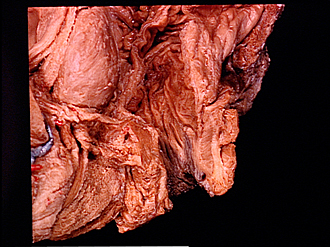
| 1
.
| Circular layer of muscular tunic of rectum |
| 2
.
| Prostate |
| 3
.
| Vesical sphincter muscle |
| 4
.
| Sphincter muscle of urethra (dissected) |
| 5
.
| Urogenital diaphragm (pointers on superior and inferior fascial layers) |
| 6
.
| Spongy part of urethra |
| 7
.
| Bulb of penis (dissected) |
| 8
.
| Coccygeus muscle |
| 9
.
| Longitudinal layer of muscular tunic of rectum |
| 10
.
| Mucosal tunic of rectum (pointer at level of perineal flexure) |
| 11
.
| Levator ani muscle (divided near its insertion into wall of anal canal) |
| 12
.
| External anal sphincter muscle |
| 13
.
| Aponeurosis of insertion of Levator ani muscle |
| 14
.
| Anal sinus |
| 15
.
| Internal anal sphincter muscle |
| 16
.
| External anal sphincter muscle (superficial part) |
| 17
.
| Level of pectinate line (not visible unless mucosa stretched) |
| 18
.
| External anal sphincter muscle (subcutaneous part) |
| 19
.
| Hemorrhoidal venous plexus (section passes through thrombus) |
| 20
.
| Anal columns |
| 21
.
| Anal verge |
| 22
.
| External anal sphincter muscle (divided see labels numbered 12, 16, 18) |
| 23
.
| Bulbospongiosus muscle (sectioned along midline) |