Bassett Collection of Stereoscopic Images of Human Anatomy
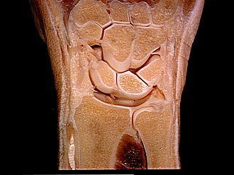
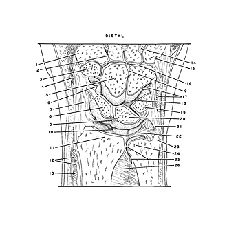
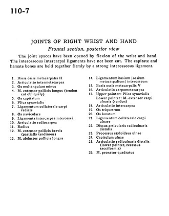
Joints of right wrist and hand
Frontal section, posterior view
Image #110-7
KEYWORDS: Fascia ligaments and tendons, Hand and fingers, Muscles and tendons, Wrist.
Creative Commons
Stanford holds the copyright to the David L. Bassett anatomical images and has assigned Creative Commons license Attribution-Share Alike 4.0 International to all of the images.

For additional information regarding use and permissions, please contact Dr. Drew Bourn at dbourn@stanford.edu.
|
| ||||||||||||||||||||||||||||||||||||||||||||||||||||||||
|
|