Bassett Collection of Stereoscopic Images of Human Anatomy
Collection Home
- Abdomen See All
- Overview 33
- Adrenal Gland 29
- Bones Cartilage Joints 4
- Central Nervous System 2
- Fascia 16
- Gallbladder 27
- Kidney 29
- Large Intestine 14
- Liver 29
- Lymphatics 12
- Muscles & Tendons 55
- Pancreas 27
- Peripheral Nervous System 56
- Small Intestine 11
- Spleen 29
- Stomach 15
- Vasculature 70
- Back See All
- Overview 5
- Bones Joints Cartilage 34
- Central Nervous System 22
- Cervical Region 42
- Lumbar Region 52
- Muscles & Tendons 40
- Meninges 5
- Peripheral Nervous System 6
- Sacral Region 37
- Thoracic Region 55
- Vasculature 17
- Vertebral Column 107
- Head See All
- Overview 49
- Bones Cartilage Joints 176
- Brain 225
- Cerebellum 51
- Cheek 41
- Connective Tissue 49
- Diencephalon 71
- Ear 37
- Exocrine & Endocrine 15
- Eye 62
- Face 153
- Frontal Lobe 22
- Medulla 32
- Meninges 23
- Midbrain 54
- Mouth 63
- Muscles & Tendons 70
- Nose 20
- Occipital Lobe 21
- Parietal Lobe 20
- Peripheral Nervous System 126
- Pons 37
- Scalp 16
- Telencephalon 134
- Temporal Lobe 28
- Vasculature 131
- Ventricules 61
- Female Pelvis See All
- Overview 1
- Anal Canal 6
- Bones Joints Cartilage 48
- Central Nervous System 26
- External Genitalia 9
- Large Intestine 6
- Muscles& Tendons 57
- Ovary 18
- Perineum 11
- Peripheral Nervous System 20
- Urinary Tract 14
- Uterus 16
- Vagina 14
- Vasculature 40
- Lower Extremity See All
- Ankle 42
- Bones Joints Cartilage 75
- Fascia 19
- Foot & Toes 78
- Knee 22
- Leg 41
- Muscles & Tendons 152
- Peripheral Nervous System 80
- Thigh 58
- Vasculature 53
- Male Pelvis See All
- Anal Canal 2
- Bones Joints Cartilage 33
- Central Nervous System 10
- Large Intestine 2
- Muscles & Tendons 37
- Perineum 3
- Peripheral Nervous System 14
- Prostate 2
- Urinary Tract 6
- Vasculature 33
- Neck See All
- Overview 13
- Bones Cartilage Joints 35
- Central Nervous System 9
- Cervical Vertebrae 23
- Esophagus 5
- Exocrine & Endocrine 20
- Fascia & Connective Tissue 37
- Lymphatics 9
- Meninges 5
- Muscles & Tendons 54
- Peripheral Nervous System 55
- Pharynx 17
- Throat 51
- Trachea 2
- Vasculature 47
- Pelvis See All
- Overview 2
- Anal Canal 6
- Bones Joints Cartilage 44
- Central Nervous System 27
- External Genitalia 10
- Female 1
- Large Intestine 6
- Muscles & Tendons 57
- Ovary 18
- Perineum 10
- Peripheral Nervous System 20
- Urinary Tract 14
- Uterus 16
- Vagina 14
- Vasculature 46
- Thorax See All
- Overview 7
- Bones Joints Cartilage 33
- Breast 7
- Central Nervous System 6
- Diaphragm 8
- Esophagus 10
- Fascia & Connective Tissue 12
- Heart 46
- Left Heart 33
- Left Lung 21
- Lung 39
- Lymphatics 9
- Mediastinum 23
- Muscles & Tendons 28
- Pericardial Sac 25
- Peripheral Nervous System 32
- Pleura 11
- Rib Cage 16
- Right Heart 31
- Right Lung 17
- Skin 2
- Thymus 2
- Vasculature 43
- Vertebral Column 20
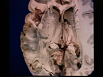
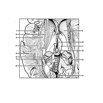
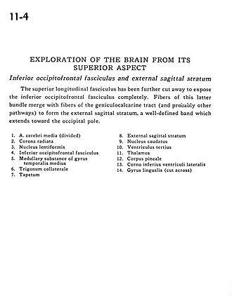
Exploration of the brain from its superior aspect
Inferior occipitofrontal fasciculus and external sagittal stratum
Image #11-4
KEYWORDS: Brain, Frontal lobe, Telencephalon, Temporal lobe.
Creative Commons
Stanford holds the copyright to the David L. Bassett anatomical images and has assigned Creative Commons license Attribution-Share Alike 4.0 International to all of the images.

For additional information regarding use and permissions, please contact Dr. Drew Bourn at dbourn@stanford.edu.
|
| ||||||||||||||||||||||||||||||||
|
|