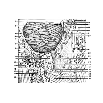
| 1
.
| Transverse sinus (opened) |
| 2
.
| Inferior cerebellar vein (lateral posterior cerebellar vein) |
| 3
.
| Dura mater |
| 4
.
| Mastoid part of temporal bone |
| 5
.
| Cerebellum (arachnoid intact over its surface) |
| 6
.
| Mastoid emissary |
| 7
.
| Occipital bone |
| 8
.
| Obliquus capitis superior muscle (cut across) |
| 9
.
| Posterior atlanto-occipital membrane |
| 10
.
| Suboccipital nerve |
| 11
.
| Occipitalis vein |
| 12
.
| Posterior arch of atlas (cut across) |
| 13
.
| Splenius capitis muscle (reflected laterally) |
| 14
.
| Semispinalis capitis muscle (cut across) |
| 15
.
| Occipitalis major nerve |
| 16
.
| Internal plate of occipital bone |
| 17
.
| Occipital diploic vein |
| 18
.
| Transverse nuchal muscle (tendinous) |
| 19
.
| External plate of occipital bone |
| 20
.
| Semispinalis capitis muscle |
| 21
.
| Subcutaneous branch of occipital artery |
| 22
.
| Occipital bone near margin of foramen magnum |
| 23
.
| Cerebellar tonsil lying deep to arachnoid membrane (Note: this extends downward slightly more than usual) |
| 24
.
| Arachnoid (overlying cerebellomedulary cistern) |
| 25
.
| Trapezius muscle |
| 26
.
| Vertebral artery (visible deep to venous plexus which surrounds it in the vertebral artery sulcus) |
| 27
.
| Posterior vertebral venous plexus |
| 28
.
| Dura mater (cut edge) |
| 29
.
| Ligamentum nuchae (cervical muscles removed from its left side) |
| 30
.
| Rectus capitis posterior major muscle (cut across near its origin from the spine of the axis) |
| 31
.
| Inferior obliquus capitis muscle |