Bassett Collection of Stereoscopic Images of Human Anatomy
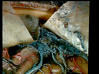
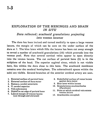
Exploration of the meninges and brain in situ
Dura reflected; arachnoid granulations projecting into venous lacunae
Image #1-3
KEYWORDS: Bones cartilage joints, Meninges, Vasculature.
Creative Commons
Stanford holds the copyright to the David L. Bassett anatomical images and has assigned Creative Commons license Attribution-Share Alike 4.0 International to all of the images.

For additional information regarding use and permissions, please contact Dr. Drew Bourn at dbourn@stanford.edu.
|

| ||||||||||||||||||||||||||||
|
|